


Overview
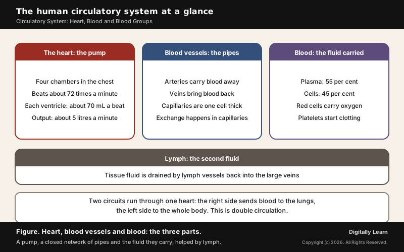
The circulatory system is the body's transport network: the heart pumps blood through a closed network of arteries, veins and capillaries, carrying oxygen and food to every cell and taking carbon dioxide and wastes away. In humans it runs as a double circulation, one circuit through the lungs and one through the body.
Human Circulatory System: Blood, Heart and Blood Vessels
Arteries, Veins and Capillaries: The Three Types of Blood Vessels
The circulatory system, also called the blood vascular system, has three parts: a muscular chambered heart, a closed network of branching blood vessels, and blood. Of the three, the vessels are the part that reaches every tissue, so they are the place to start.
Blood leaves the heart under high pressure and returns to it under very little, so the vessels on the two journeys are built differently. The walls of arteries and veins have the same three layers: an inner lining of flat cells called the tunica intima, a middle layer of smooth muscle and elastic fibres called the tunica media, and an outer coat of fibrous connective tissue called the tunica externa. The middle layer is much thinner in veins.
| Vessel | What it does | How it is built |
|---|---|---|
| Arteries | Carry blood away from the heart to the organs | Thick, elastic walls that withstand high pressure |
| Veins | Bring blood back from the organs to the heart | Thinner walls with valves that keep blood flowing one way |
| Capillaries | Exchange materials with the surrounding cells | Walls only one cell thick |
An artery divides into ever smaller vessels until it reaches the capillaries, where food, oxygen and wastes cross between blood and cells. The capillaries then join to form veins. Because veins are wide and stretchable, they hold most of the blood at any moment: nearly 70 per cent of the body's blood is in the veins.
Open and Closed Circulation and the Two-, Three- and Four-Chambered Heart
Animals move blood in one of two ways. In an open circulatory system, found in arthropods and molluscs, the heart pumps blood through large vessels into open spaces in the body called sinuses. In a closed circulatory system, found in annelids and chordates, blood always stays inside a network of vessels. The closed pattern is the more advantageous one because the flow can be regulated precisely.
- Fishes: A two-chambered heart with one atrium and one ventricle. The heart pumps deoxygenated blood to the gills, and oxygenated blood goes straight on to the body, so blood passes through the heart only once in each round: single circulation.
- Amphibians and most reptiles: A three-chambered heart with two atria and one ventricle. Oxygenated and deoxygenated blood mix in the single ventricle: incomplete double circulation.
- Crocodiles, birds and mammals: A four-chambered heart with two atria and two ventricles. The two kinds of blood never mix: double circulation.
Keeping oxygen-rich blood apart from oxygen-poor blood gives a highly efficient oxygen supply. Birds and mammals need it, because they spend a great deal of energy keeping their body temperature constant; animals whose body temperature follows their surroundings can tolerate some mixing.
Composition of Blood: Plasma and Formed Elements
Plasma Proteins and the Difference Between Blood Plasma and Serum
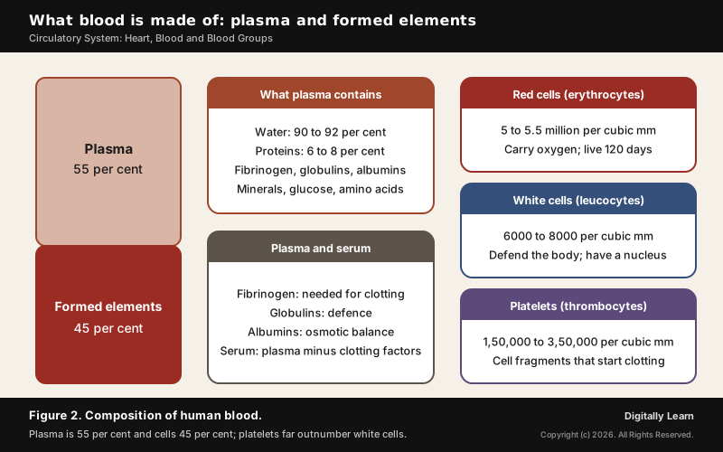
Blood is a special connective tissue made of a fluid matrix, the plasma, in which the formed elements (the cells and cell fragments) float. It counts as a connective tissue because it is made of cells held in a matrix, here a fluid one. Normal blood is slightly alkaline, with a pH of 7.35 to 7.45.
Plasma is a straw-coloured, sticky fluid that makes up nearly 55 per cent of blood. It is 90 to 92 per cent water and 6 to 8 per cent plasma proteins, and it also carries minerals such as sodium, calcium, magnesium, bicarbonate and chloride, along with glucose, amino acids and lipids in transit. Plasma transports food, carbon dioxide and nitrogenous wastes in dissolved form.
- Fibrinogen: Needed for the clotting of blood.
- Globulins: Mainly involved in the body’s defence.
- Albumins: Help keep the osmotic balance between blood and tissues.
Red Blood Cells and Haemoglobin: Oxygen Carriers and Anaemia
Red blood cells, or erythrocytes, are the most numerous cells in blood: a healthy adult man has on average 5 to 5.5 million of them in each cubic millimetre. In adults they are made in the red bone marrow. In most mammals they have no nucleus and are biconcave discs.
Red cells carry haemoglobin, an iron-containing protein that is red, which is why blood is red. A healthy person has 12 to 16 grams of haemoglobin in every 100 millilitres of blood, and it is haemoglobin that carries most of the oxygen from the lungs to the tissues. A red cell lives about 120 days, after which it is destroyed in the spleen, often called the graveyard of red cells.
Anaemia is an insufficient mass of red cells. It can result from bleeding, from blood disorders such as thalassaemia, or from nutritional deficiencies; iron deficiency and India's anaemia programme are covered in vitamins, minerals and malnutrition. An inherited cause is sickle cell disease, in which faulty haemoglobin makes red cells take a sickle shape; such cells cannot bend through capillaries and block them. The disease is found in tribal regions of India; stem cells and blood disorders are taken up in the cell cycle and stem cells.
White Blood Cells and Platelets: Types and Functions
White blood cells, or leucocytes, have no haemoglobin and so are colourless. They have a nucleus, live only a short time, and number 6000 to 8000 in each cubic millimetre of blood. They fall into two groups: granulocytes (neutrophils, eosinophils and basophils) and agranulocytes (lymphocytes and monocytes).
| Type | Share of white cells | Main job |
|---|---|---|
| Neutrophils | 60 to 65 per cent, the most numerous | Engulf and destroy invading organisms |
| Eosinophils | 2 to 3 per cent | Resist infections; linked with allergic reactions |
| Basophils | 0.5 to 1 per cent, the fewest | Release histamine, serotonin and heparin in inflammation |
| Lymphocytes | 20 to 25 per cent | B and T lymphocytes carry out immune responses |
| Monocytes | 6 to 8 per cent | Engulf and destroy invading organisms |
Platelets, or thrombocytes, are not whole cells but fragments of large bone-marrow cells called megakaryocytes. Blood normally holds 1,50,000 to 3,50,000 of them per cubic millimetre, far more than white cells. Platelets release most of the substances that start clotting, so a fall in their number leads to clotting disorders and heavy loss of blood.
Blood Groups: ABO System, Rh Factor and Safe Blood Transfusion
ABO Blood Groups Chart: Universal Donor and Universal Recipient
Blood groups sort people by the antigens on the surface of their red cells. Karl Landsteiner described the ABO system in 1901, and ABO and Rh are the two groupings used all over the world. In the ABO system two surface antigens, A and B, decide the group, and the plasma carries natural antibodies against whichever antigen the cells lack.
| Group | Antigen and antibody | In a transfusion |
|---|---|---|
| A | Antigen A; anti-B antibody | Its anti-B attacks any red cells carrying B |
| B | Antigen B; anti-A antibody | Its anti-A attacks any red cells carrying A |
| AB | Antigens A and B; no antibody | Nothing in its plasma attacks A or B cells |
| O | No antigen; anti-A and anti-B antibodies | Its cells carry nothing for anti-A or anti-B to attack |
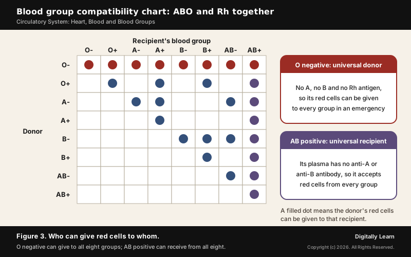
Blood must be matched before a transfusion, because a recipient's antibodies attack donor cells carrying the matching antigen and make them clump. Group O is the universal donor: its red cells carry neither A nor B, so no recipient's antibody attacks them. Group AB is the universal recipient: its plasma has no anti-A or anti-B antibody, so it accepts red cells from every group. When the Rh antigen is counted too, O negative red cells can go to all eight groups, which is why O negative blood is used when a transfusion cannot wait for the patient's group to be tested. The blood groups chart below shows which of the eight groups can give red cells to which.
Blood groups are inherited. The ABO group is controlled by one gene, I, with three alleles: IA and IB each make a form of sugar on the red cell, while i makes none. IA and IB are both dominant over i, and when they occur together both are expressed, an example of co-dominance. A child therefore takes one allele from each parent: an AB parent passes on IA or IB, and an O parent can pass on only i, so their children can be group A or group B but never O or AB.
Rh Factor and Erythroblastosis Foetalis
The Rh factor is a second antigen on red cells, named after a similar antigen in Rhesus monkeys. Nearly 80 per cent of people carry it and are Rh positive; the rest are Rh negative. Landsteiner and Wiener reported it in 1940. An Rh negative person exposed to Rh positive blood forms antibodies against it, so the Rh group must also be matched before a transfusion.
- First pregnancy: An Rh negative mother carrying an Rh positive foetus is usually safe, because the placenta keeps the two bloods apart.
- At delivery: Small amounts of the baby’s Rh positive blood can reach the mother, who then starts making anti-Rh antibodies.
- Next pregnancy: These antibodies can cross into an Rh positive foetus and destroy its red cells, causing severe anaemia and jaundice or even death. This condition is erythroblastosis foetalis.
- Prevention: Anti-Rh antibodies are given to the mother immediately after the first delivery.
Rare Blood Groups in India: Bombay Blood Group and Blood Donation
The Bombay blood group (hh) is one of the rare blood groups in India and among the rarest blood types anywhere. It was first found in Bombay by Dr. Yeshwant Madhav Bhende in 1952 and occurs mostly in the Indian subcontinent and Iran. People with it lack the H antigen, the base substance from which A and B antigens are made, so their cells carry none of the three. Ordinary ABO tests show them as group O, but they can receive blood only from another Bombay donor.
The Bombay phenotype is found in about 4 people in a million worldwide, but in parts of Mumbai it can reach about 1 in 10,000. Because matching donors are so few, a person with this group who needs an urgent transfusion may be unable to get it in time.
Blood transfusion in India is governed through the National Blood Transfusion Council, the apex policy body, with a council in each state. A Supreme Court judgment of 1996 led to a complete ban on professional (paid) blood donation, and the National Blood Policy of 2002 set the framework for safe, voluntary donation. The country's annual need is estimated at 14.6 million units. The national portal e-RaktKosh lists blood availability and camps.
- Who can donate: People aged 18 to 65 who weigh 45 kg or more and are fit and healthy.
- How often: Men can donate whole blood again after 90 days and women after 120 days. After a donation the body replaces the lost blood, which stimulates the production of new blood cells.
- What one unit gives: Separated into red cells, plasma and platelets, one donated unit can help up to four patients. Red cells keep for 42 days, platelets for 5 days and frozen plasma for a year.
Blood Clotting and the Lymphatic System
Coagulation of Blood: Thrombin, Fibrin and Calcium Ions
Blood clotting, or coagulation, stops a wound from bleeding for long. The dark reddish-brown scab over a cut is a clot: a network of threads called fibrin in which dead and damaged blood cells are trapped. The clot forms through a chain of reactions.
- Trigger: An injury makes platelets release certain factors; the injured tissue releases factors of its own.
- Cascade: A series of linked enzyme reactions activates clotting factors that were present in the plasma in an inactive state, producing an enzyme complex called thrombokinase.
- Thrombin: Thrombokinase converts inactive prothrombin in the plasma into the enzyme thrombin.
- Fibrin threads: Thrombin converts soluble fibrinogen into insoluble fibrin threads, which form the clot.
Calcium ions play a very important part in this chain. When one of the clotting factors is missing, blood fails to clot properly. In haemophilia, an inherited disorder, clotting factor VIII (haemophilia A) or factor IX (haemophilia B) is low. The genes lie on the X chromosome, so the disease is far more common in males; India's framework for it is covered in haemophilia and rare diseases in India.
Lymph, Tissue Fluid and Lymphocytes
As blood passes through the capillaries, some water and small dissolved substances leak out into the spaces between cells, leaving the large proteins and most blood cells behind. This tissue fluid has the same minerals as plasma, and every exchange of nutrients and gases between blood and cells passes through it.
A separate network of vessels, the lymphatic system, collects this fluid and drains it back into the large veins. Once inside the lymph vessels the fluid is called lymph: colourless, with less protein than plasma, and carrying specialised lymphocytes for the body's immune responses. Lymph also carries nutrients and hormones, and the fat absorbed from food enters it through the lacteals in the villi of the small intestine, described in the human digestive system.
| Feature | Blood | Lymph |
|---|---|---|
| Colour | Red, from haemoglobin in red cells | Colourless |
| Proteins | Plasma rich in proteins | Fewer proteins than plasma |
| Cells | Red cells, white cells and platelets | Mainly lymphocytes |
| Flow | Pumped round by the heart in a closed circuit | Drains one way from tissues into the veins |
Structure and Working of the Human Heart
Chambers, Valves and Coronary Arteries of the Heart
The human heart sits in the chest cavity between the two lungs, slightly tilted to the left, and is about the size of a clenched fist. A double-walled bag called the pericardium, filled with pericardial fluid, protects it. The heart has four chambers: two small upper atria that receive blood and two larger lower ventricles that pump it out.
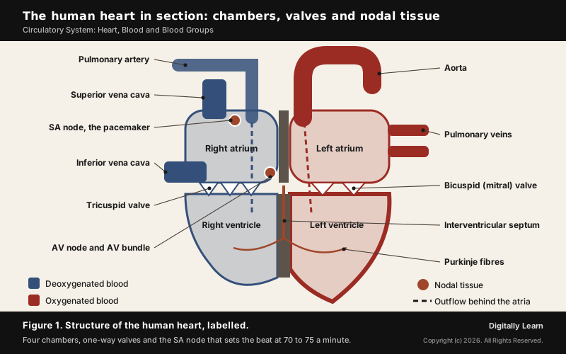
- Septa: A thin inter-atrial septum separates the two atria and a thick inter-ventricular septum separates the two ventricles, so the right and left sides never mix.
- Tricuspid valve: Three flaps guard the opening between the right atrium and right ventricle.
- Bicuspid or mitral valve: Two flaps guard the opening between the left atrium and left ventricle.
- Semilunar valves: These guard the exits from the ventricles into the pulmonary artery and the aorta. All the valves let blood flow one way only and stop it flowing back.
The whole heart is made of cardiac muscle, and the ventricles have much thicker walls than the atria because they must pump blood out to the lungs and the rest of the body. The heart muscle itself needs a blood supply of its own: a special coronary system of vessels carries blood to and from the cardiac muscle, and the arteries that supply the heart are the coronary arteries.
SA Node and AV Node: The Heart's Natural Pacemaker
The heart starts each beat by itself, without needing a signal from the brain. A specialised kind of cardiac muscle, the nodal tissue, can generate electrical impulses by itself; it is auto-excitable. Because the heart's rhythm starts in its own muscle, the human heart is called myogenic.
- Sino-atrial node (SAN): A patch in the upper right corner of the right atrium.
- Atrio-ventricular node (AVN): A mass in the lower left corner of the right atrium, next to the septum between atrium and ventricle.
- AV bundle and bundle of His: Nodal fibres that run from the AVN through the septum and divide into right and left branches.
- Purkinje fibres: Fine fibres that carry the impulse through the muscle of each ventricle.
Different parts of this system can fire at different rates, but the SAN fires fastest, at 70 to 75 times a minute. It therefore starts and keeps the rhythm of every beat, which is why the SA node is called the pacemaker. The normal heart beats 70 to 75 times a minute, 72 on average.
Cardiac Cycle, Heart Sounds and Cardiac Output
The cardiac cycle is the sequence of contraction (systole) and relaxation (diastole) of the atria and ventricles that repeats with every beat. It runs in a fixed order.
- Joint diastole: All four chambers are relaxed. Blood from the veins flows through the atria into the ventricles, because the tricuspid and bicuspid valves are open.
- Atrial systole: The SAN fires and both atria contract together, which raises the flow of blood into the ventricles by about 30 per cent.
- Ventricular systole: The impulse passes through the AVN and bundle of His and the ventricles contract. The rising pressure shuts the tricuspid and bicuspid valves and forces open the semilunar valves, sending blood into the pulmonary artery and aorta.
- Ventricular diastole: The ventricles relax, the semilunar valves close to stop backflow, and the tricuspid and bicuspid valves open again as blood arrives from the atria.
At 72 beats a minute each cycle lasts about 0.8 seconds. In each beat every ventricle pumps out about 70 millilitres, the stroke volume. Stroke volume multiplied by heart rate gives the cardiac output, about 5 litres a minute in a healthy person; an athlete's cardiac output is much higher, because the body can raise both numbers.
The closing valves make the two heart sounds a doctor hears through a stethoscope. The first sound, lub, comes from the closing of the tricuspid and bicuspid valves; the second, dub, from the closing of the semilunar valves.
Electrocardiogram (ECG): P Wave, QRS Complex and T Wave
An electrocardiogram, or ECG, is a graph of the heart's electrical activity during the cardiac cycle, recorded by a machine called an electrocardiograph. For a standard ECG the patient is connected by three leads, one to each wrist and one to the left ankle; a detailed evaluation attaches more leads to the chest, as in the 12-lead ECG. Willem Einthoven's string galvanometer of 1903 made precise recording possible, and it won him the Nobel Prize in 1924.
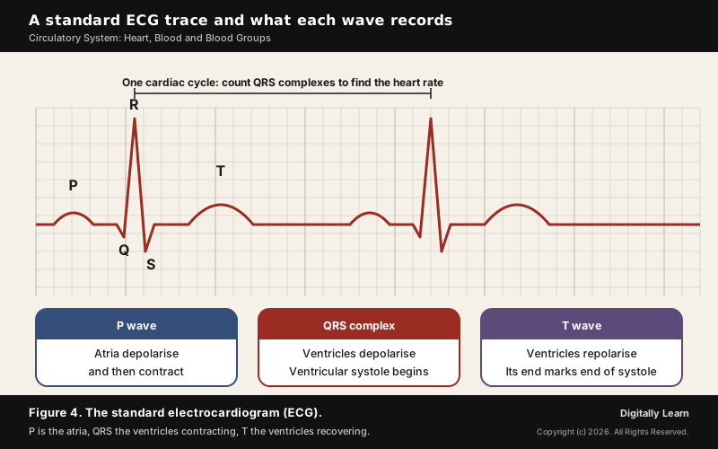
- P wave: The electrical excitation, or depolarisation, of the atria, which leads to their contraction.
- QRS complex: The depolarisation of the ventricles, which starts their contraction; systole begins shortly after Q.
- T wave: The return of the ventricles to their resting state, or repolarisation; the end of the T wave marks the end of systole.
Counting the QRS complexes in a given time gives the heart rate. Because ECGs from different people have roughly the same shape for a given set of leads, any departure from that shape points to a possible abnormality, which is what makes the ECG so useful in diagnosis.
Double Circulation and Regulation of the Heart
Pulmonary and Systemic Circulation, Hepatic Portal and Coronary Systems
Double circulation means that blood passes through the heart twice in each complete round of the body, along two separate circuits that never mix.
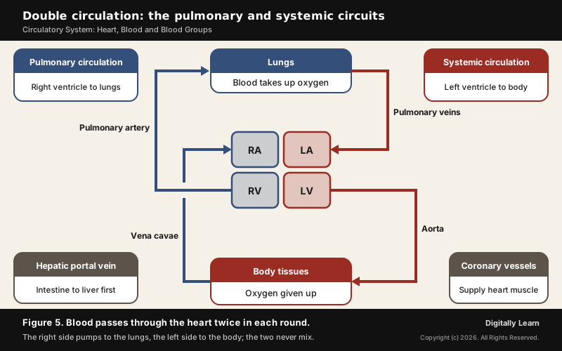
- Pulmonary circulation: The right ventricle pumps deoxygenated blood into the pulmonary artery and on to the lungs. The oxygenated blood returns through the pulmonary veins to the left atrium.
- Systemic circulation: The left ventricle pumps oxygenated blood into the aorta, and a network of arteries, arterioles and capillaries carries it to the tissues. Venules, veins and the vena cavae collect the deoxygenated blood and empty it into the right atrium.
Two special routes sit within the system. The hepatic portal system is a unique link between the digestive tract and the liver: the hepatic portal vein carries blood from the intestine to the liver before it enters the systemic circulation. The coronary system serves only the heart muscle. Because the two circuits never mix, the blood the aorta sends to the body always carries its full load of oxygen.
Nervous and Hormonal Control of Heart Rate and Blood Vessels
The heart sets its own basic rhythm, but the body can speed it up or slow it down. A special centre in the medulla oblongata of the brain adjusts the heart through the autonomic nervous system.
- Sympathetic nerves: Raise the heart rate and the force of ventricular contraction, and so the cardiac output.
- Parasympathetic nerves: Lower the heart rate and the speed at which the impulse is conducted, and so the cardiac output.
- Adrenal medullary hormones: Also increase the cardiac output.
Blood flow also depends on the width of the vessels. The inner lining of blood vessels makes nitric oxide, a gas that signals the surrounding smooth muscle to relax. The vessel widens, which is called vasodilation, and more blood flows through it. Narrowing of the small arteries has the opposite effect and raises blood pressure.
Disorders of the Circulatory System
Blood Pressure and Hypertension: Normal Values and Measurement
Blood pressure is the force that blood exerts against the wall of a vessel. It is much higher in arteries than in veins. It is written as two numbers: the systolic pressure, while the ventricles contract, and the diastolic pressure, while they relax. The normal value is about 120/80 millimetres of mercury, and blood pressure is measured with an instrument called a sphygmomanometer.
Hypertension, or high blood pressure, is diagnosed when repeated readings are 140/90 or higher. It is caused by the narrowing of the small arteries, the arterioles, which raises the resistance to blood flow; it can burst an artery and cause internal bleeding, and it leads to heart disease and damages the brain and kidneys. Most people with hypertension feel no symptoms, so the only way to know is to have the blood pressure checked.
- Worldwide: An estimated 1.4 billion adults aged 30 to 79 have hypertension; about 600 million of them (44 per cent) are unaware of it, and only about 320 million (23 per cent) have it under control.
- India’s screening: Under the National Programme for Prevention and Control of Non-Communicable Diseases, everyone over 30 is to be screened; as of February 2026 over 40.87 crore people had been screened for hypertension, 7 crore diagnosed and 5.57 crore were under treatment.
- The 75/25 initiative: Announced in 2023, it aimed to put 75 million people with hypertension and diabetes on standard care through primary health centres by 2025.
Coronary Artery Disease, Angina and Heart Failure
Cardiovascular diseases are the leading cause of death in the world: an estimated 19.8 million people died from them in 2022, about 32 per cent of all deaths, and 85 per cent of these deaths were due to heart attack and stroke. Three conditions of the heart are named separately.
- Coronary artery disease: Often called atherosclerosis, it narrows the vessels that supply the heart muscle, through deposits of calcium, fat, cholesterol and fibrous tissue.
- Angina (angina pectoris): Acute chest pain when not enough oxygen reaches the heart muscle; it is more common in middle-aged and elderly people.
- Heart failure: The heart does not pump blood well enough to meet the body’s needs. It is also called congestive heart failure, because congestion of the lungs is one of its main signs.
The main behavioural risks for heart disease and stroke are an unhealthy diet, physical inactivity, tobacco use and the harmful use of alcohol; air pollution is an important environmental risk. How the lungs load the blood with oxygen in the first place is explained in the human respiratory system.
Previous Year UPSC-CSE Questions
Previous Year UPSC-CSE Questions By the end you will be able to draft model answers for the following UPSC questions. Each question carries a collapsible framework showing how to approach it in the exam.
- UPSC Prelims 2024 Prelims-GSWhich one of the following is synthesised in human body that dilates blood vessels and increases blood flow ?
How to approach this Prelims question
Approach: Four oxides of nitrogen with similar names; ask which one the body itself makes as a signal to vessel muscle.
Trap to watch: The four names differ by one word; only nitric oxide is made by the vessel lining as a signal to widen the vessel.
Key facts to recall:
- (a) Nitric oxide: right. The inner lining of vessels makes it; it relaxes the smooth muscle of the vessel wall, the vessel widens and blood flow rises.
- (b) Nitrous oxide: wrong. A different oxide of nitrogen; it is not the signal the vessel lining makes to widen the vessel.
- (c) Nitrogen dioxide: wrong. Also a different oxide of nitrogen, not a signalling molecule made by the vessel wall.
- (d) Nitrogen pentoxide: wrong. Another oxide of nitrogen with no role as the body's vessel-widening signal.
Answer signal: Nitric oxide, so option (a) is the answer.
- UPSC Prelims 2011 Prelims-GSA married couple adopted a male child. A few years later twin boys were born to them. The blood group of the couple is AB positive and O negative. The blood group of the three sons is A positive, B positive, and O positive. The blood group of the adopted son is
How to approach this Prelims question
Approach: Work out which ABO groups the two parents can produce. The AB parent passes on IA or IB; the O parent passes on i. So every biological child is group A (IAi) or group B (IBi), never O. The son whose group falls outside that set is the adopted one. The Rh signs do not change this.
Trap to watch: The Rh signs are a distraction: the ABO letters alone settle the question.
Key facts to recall:
- (a) O positive: right. Group O needs i from both parents, and the AB parent has no i allele to give, so this son cannot be their biological child.
- (b) A positive: wrong. An A child (IAi) can be born to AB and O parents, so this son can be biological.
- (c) B positive: wrong. A B child (IBi) can also be born to these parents.
- (d) Cannot be determined: wrong. The ABO alleles alone settle which son is adopted.
Answer signal: The O positive son cannot be their biological child, so option (a) is the answer.
- UPSC Prelims 2008 Prelims-GSWhat is the pH level of blood of a normal person?
How to approach this Prelims question
Approach: Blood is slightly alkaline, so the answer lies just above 7.
Trap to watch: Option (b) is slightly acidic and option (d) is far too alkaline; only one range sits just above neutral.
Key facts to recall:
- (a) 4.5 to 4.6: wrong. Strongly acidic, far below normal blood.
- (b) 6.45 to 6.55: wrong. Slightly acidic; blood is slightly alkaline.
- (c) 7.35 to 7.45: right. Normal blood pH; below 7.35 blood is too acidic and above 7.45 too alkaline.
- (d) 8.25 to 8.35: wrong. Far too alkaline for normal blood.
Answer signal: 7.35 to 7.45, so option (c) is the answer.
- UPSC Prelims 2004 Prelims-GSIn which organ of the human body are the lymphocyte cells formed?
How to approach this Prelims question
Approach: Ask where all blood cells are made: the bone marrow. Then find the option that contains bone marrow.
Trap to watch: The spleen is linked with red cells being destroyed and with lymph tissue, which makes it tempting; formation happens in the marrow.
Key facts to recall:
- (a) Liver: wrong. Blood cells, lymphocytes included, are formed in the bone marrow, not the liver.
- (b) Long bone: right. Bone marrow produces red cells, white cells and platelets, and in adults red marrow lies in flat bones and the ends of long bones.
- (c) Pancreas: wrong. It is a composite gland that makes digestive juice and hormones such as insulin, not blood cells.
- (d) Spleen: wrong. It is a secondary lymphoid organ where mature lymphocytes meet antigens, and it destroys old red cells; lymphocytes are formed in the marrow.
Answer signal: The long bone, which holds the marrow, so option (b) is the answer.
- UPSC Prelims 2002 Prelims-GSWith reference to the blood in a normal person, which one of the following statements is correct?
How to approach this Prelims question
Approach: Test each option against one number: the share of blood in veins, the share of cells, where white cells are made, and the two counts.
Trap to watch: Option (b) uses the figure 70 per cent, which belongs to veins' share of blood, not to cells' share of blood volume.
Key facts to recall:
- (a) Veins less numerous and holding less blood: wrong. Veins hold nearly 70 per cent of the blood at any time.
- (b) Blood cells about 70 per cent of blood volume: wrong. Formed elements are about 45 per cent; the 70 per cent figure belongs to the veins' share of blood.
- (c) White cells made by lymph nodes only: wrong. White cells are made in the bone marrow.
- (d) More platelets than white cells: right. Platelets number 1,50,000 to 3,50,000 per cubic mm against 6,000 to 8,000 white cells.
Answer signal: Only option (d) is correct.
- UPSC Prelims 2001 Prelims-GSA man whose blood group is not known meets with a serious accident and needs blood transfusion immediately. Which one of the blood groups mentioned below and readily available in the hospital will be safe for transfusion?
How to approach this Prelims question
Approach: Pick the group whose red cells carry no antigen that any recipient could attack.
Trap to watch: O positive still carries the Rh antigen and could harm an Rh negative patient.
Key facts to recall:
- (a) O, Rh negative: right. Its red cells carry no A, no B and no Rh antigen, so no recipient's antibodies can attack them.
- (b) O, Rh positive: wrong. The cells carry the Rh antigen and could harm an Rh negative patient.
- (c) AB, Rh negative: wrong. The cells carry A and B antigens, which the anti-A or anti-B of group A, B or O patients would attack.
- (d) AB, Rh positive: wrong. It carries A, B and Rh antigens, the least safe choice of all.
Answer signal: O negative, so option (a) is the answer.
- UPSC Prelims 1997 Prelims-GSArteries supplying blood to the heart are called
How to approach this Prelims question
Approach: Match each artery to the organ it serves.
Trap to watch: The pulmonary arteries leave the heart for the lungs; they do not supply the heart muscle.
Key facts to recall:
- (a) Carotid arteries: wrong. They run up the neck and supply the brain.
- (b) Hepatic arteries: wrong. They supply the liver.
- (c) Coronary arteries: right. They supply the heart muscle itself.
- (d) Pulmonary arteries: wrong. They leave the heart for the lungs; they do not supply the heart muscle.
Answer signal: Coronary arteries, so option (c) is the answer.
- UPSC Prelims 1995 Prelims-GSA person with ‘AB’ blood group is sometimes called a universal recipient because of the
How to approach this Prelims question
Approach: Recall what the AB group has on its cells and what it has in its plasma.
Trap to watch: Group AB has two antigens, not none; the key is the missing antibodies.
Key facts to recall:
- (a) Lack of antigen: wrong. AB red cells carry both antigen A and antigen B.
- (b) Lack of antibodies: right. AB plasma has neither anti-A nor anti-B, so it attacks no donor's cells.
- (c) Lack of both antigens and antibodies: wrong. AB lacks antibodies but carries two antigens.
- (d) Presence of antibodies: wrong. It is the absence of antibodies that makes AB the universal recipient.
Answer signal: Lack of antibodies, so option (b) is the answer.
Sources and Further Reading
- NCERT: Biology, Class 11, Chapter 15, Body Fluids and Circulation
- NCERT: Science, Class 10, Chapter 5, Life Processes
- NCERT: Biology, Class 12, Chapter 4, Principles of Inheritance and Variation
- World Health Organization: Hypertension, fact sheet
- World Health Organization: Cardiovascular diseases, fact sheet
- Press Information Bureau: launch of the National Sickle Cell Anaemia Elimination Mission, 1 July 2023
- Press Information Bureau: the 75/25 initiative on hypertension and diabetes, 2023
- Press Information Bureau: update on NP-NCD screening, 23 March 2026
- Directorate General of Health Services: Blood Transfusion Services
- e-RaktKosh: national blood bank management portal
- Wikipedia: Blood
- Wikipedia: Blood type
- Wikipedia: Rh blood group system
- Wikipedia: Hh blood group (Bombay phenotype)
- Wikipedia: Nitric oxide
- Wikipedia: Vein
- Wikipedia: Bone marrow
- Wikipedia: Haemophilia
- Wikipedia: Sickle cell disease
- Wikipedia: Electrocardiography
Editorial Disclaimer
This article is for UPSC preparation and explains how the human circulatory system works. It is not medical advice for any individual.


