

Overview
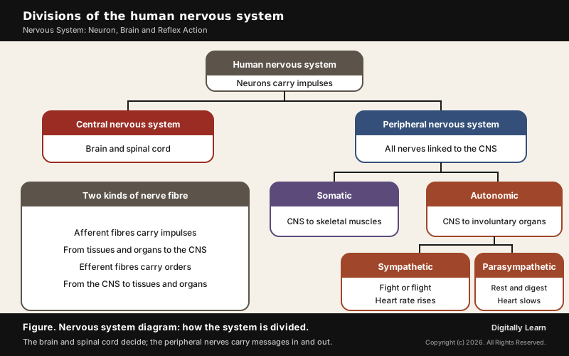
The nervous system is the body's fast control network: nerve cells called neurons carry electrical impulses between the brain, the spinal cord and every part of the body. The brain and spinal cord form the central nervous system, and the nerves linking them to the body form the peripheral nervous system.
Nervous System and Endocrine System: Two Ways the Body Coordinates
Difference Between Nervous and Hormonal Control
The organs of the body have to work in step, and keeping them so is called coordination. During exercise, for example, the muscles need more energy, so breathing, heartbeat and blood flow all rise together and return to normal afterwards. Two systems produce this coordination: the nervous system, whose specialised cells called neurons detect, receive and pass on stimuli, and the endocrine system.
Electrical signals have limits. An impulse reaches only the cells that nerves connect, and after sending one impulse a cell needs time to reset before it can send another. Hormones make up for this: they are slower, but they can reach every cell in the body, whatever its nerve connections, and they can act steadily over time.
| Feature | Nervous system | Endocrine system |
|---|---|---|
| Messenger | Nerve impulses, passed on by chemicals at synapses | Hormones carried in the blood |
| Reach | Only cells connected by nerves | Any cell in the body |
| Speed | Quick, point-to-point | Slower but steady |
The two systems work as partners, and the hypothalamus in the brain links them, since it also makes hormones. The endocrine side of the story, with each gland and hormone, is the subject of a separate article; this page follows the nervous side.
Neuron Structure, Nerve Impulse and Synapse
Neuron Diagram and Structure: Dendrites, Cell Body, Axon and Myelin Sheath
A neuron is a nerve cell, the structural and functional unit of the nervous system. It has three main parts, and each has its own job in passing an impulse along.
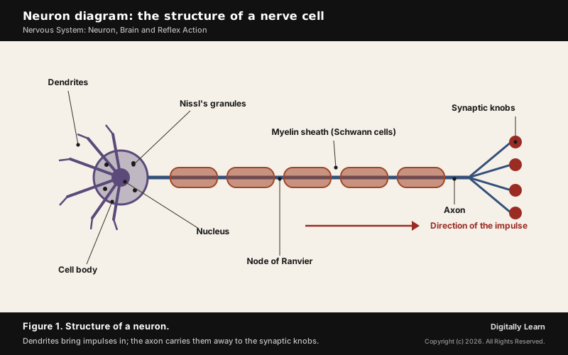
- Cell body: Holds the cytoplasm, the usual cell organelles and granular Nissl’s granules.
- Dendrites: Short, repeatedly branched fibres that carry impulses towards the cell body.
- Axon: A long fibre that carries impulses away from the cell body; its end branches into synaptic knobs holding vesicles of chemicals called neurotransmitters.
Many axons are wrapped in a myelin sheath formed by Schwann cells, with gaps called nodes of Ranvier; such myelinated fibres are found in the spinal and cranial nerves, while unmyelinated fibres are common in the autonomic and somatic systems. Neurons are also sorted by their fibres: multipolar neurons (one axon, two or more dendrites) in the cerebral cortex, bipolar neurons in the retina of the eye, and unipolar neurons, usually found in the embryo. How nerve cells fit the wider picture of cells is covered in cell structure and function.
Resting Potential and Action Potential: How a Nerve Impulse Travels
A nerve impulse is an electrical signal that runs along the membrane of an axon. Neurons can carry it because their membranes are polarised: the charge outside differs from the charge inside, and a stimulus can flip it for a moment.
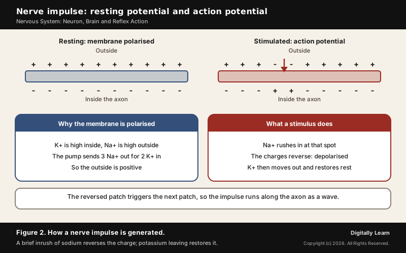
- At rest: The membrane lets potassium through but almost no sodium, so the axon holds a lot of potassium and negatively charged proteins inside, and the fluid outside holds a lot of sodium. The sodium-potassium pump keeps it so, sending 3 sodium ions out for every 2 potassium ions in. The outside is positive and the inside negative; this charge difference is the resting potential.
- Stimulus: At the stimulated spot the membrane suddenly lets sodium in. Sodium rushes in, the outside turns negative and the inside positive, and the membrane is depolarised. This reversed charge is the action potential, which is the nerve impulse.
- Spread: Current flows from the stimulated spot to the next one, reversing it in turn, so the impulse travels along the whole axon.
- Recovery: Within a fraction of a second potassium moves out, the resting potential returns, and the fibre is ready for the next stimulus.
Synapse and Neurotransmitters: Electrical and Chemical Transmission
A synapse is the junction where an impulse passes from one neuron to the next. It is formed by the membranes of a pre-synaptic and a post-synaptic neuron, which may or may not be separated by a gap called the synaptic cleft.
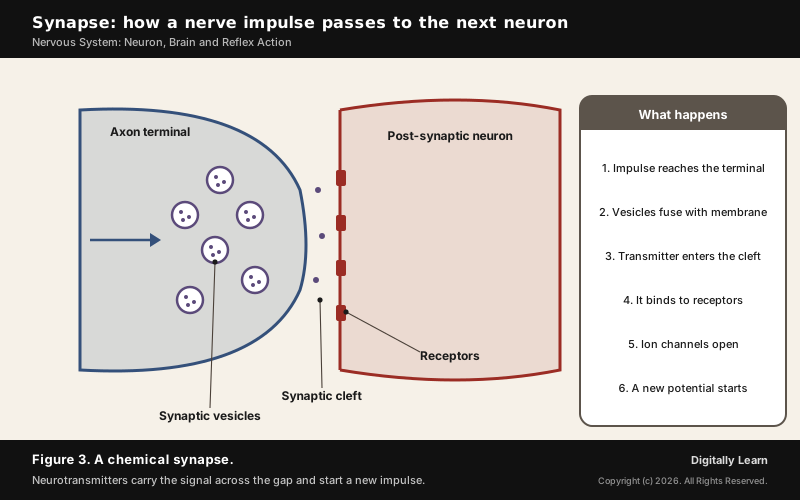
- Electrical synapse: The two membranes are very close and current flows straight across, so transmission is always faster; such synapses are rare in the human body.
- Chemical synapse: A fluid-filled synaptic cleft separates the membranes. An arriving impulse makes vesicles release neurotransmitters into the cleft; they bind to receptors on the next neuron, open ion channels and start a new potential, which may be excitatory or inhibitory.
More than 100 neurotransmitters have been identified, and whether a synapse excites or calms the next neuron depends largely on which one it uses. Four matter most for the examination.
- Glutamate: Used at the great majority of fast excitatory synapses in the brain and spinal cord.
- GABA: Used at the great majority of fast inhibitory synapses in almost every part of the brain; many sedative and tranquillising drugs work by strengthening its effect.
- Acetylcholine: After it acts, the enzyme acetylcholinesterase cuts it apart, and the choline left over is taken back by the first neuron and recycled.
- Dopamine: Its loss underlies Parkinson’s disease, and cocaine interferes with its transport, both explained later on this page.
Divisions of the Human Nervous System
Central Nervous System: Brain, Spinal Cord, Meninges and Cerebrospinal Fluid
The central nervous system (CNS) is made of the brain and the spinal cord, and it is where information is processed and from where the body is controlled. Neural organisation is very simple in lower invertebrates: in Hydra the neural system is only a network of neurons, insects have a brain along with a number of ganglia, and vertebrates have a more developed neural system still. The brain is the body's command and control system: it controls voluntary movements, balance, the working of vital involuntary organs such as the lungs, heart and kidneys, body temperature, hunger and thirst, the 24-hour body rhythms and several endocrine glands, and it is the seat of vision, hearing, speech, memory, intelligence, emotion and thought.
- Skull and meninges: The brain sits in the bony skull and is wrapped in three membranes, the cranial meninges: the outer dura mater, the very thin middle arachnoid and the inner pia mater, which touches the brain.
- Cerebrospinal fluid: Inside the skull the brain sits in a fluid-filled cushion that absorbs shocks. This cerebrospinal fluid (CSF) is made mostly by the choroid plexuses in the ventricles of the brain; about 125 mL is present at any one time and about 500 mL is made every day.
- Vertebral column: The backbone protects the spinal cord.
Peripheral and Autonomic Nervous System: Sympathetic and Parasympathetic
The peripheral nervous system (PNS) is made of all the nerves that link the CNS to the rest of the body: cranial nerves from the brain and spinal nerves from the spinal cord. Its fibres run in two directions. Afferent fibres carry impulses from tissues and organs to the CNS, and efferent fibres carry orders from the CNS back to them.
| Division | What it serves |
|---|---|
| Somatic | Skeletal muscles, as when you decide to move an arm |
| Sympathetic (autonomic) | Involuntary organs and smooth muscle; sets off fight or flight |
| Parasympathetic (autonomic) | Involuntary organs and smooth muscle; promotes rest and digestion |
The autonomic nervous system runs the organs we do not control by thought, such as the heart rate, digestion, breathing rate and the size of the pupil. Its two branches usually pull in opposite directions: the sympathetic system sets off the fight-or-flight response, while the parasympathetic system promotes rest and digestion.
| Organ | Fight or flight | Rest and digest |
|---|---|---|
| Heart | Raises the heart rate and the force of contraction | Lowers the heart rate and cardiac output |
| Airways | Widens the bronchioles for more oxygen | Narrows them when less oxygen is needed |
| Eye | Widens the pupil to let in more light | Narrows the pupil for close vision |
| Gut | Slows peristalsis | Speeds peristalsis and saliva for digestion |
Reflex Action and the Reflex Arc
A reflex action is a sudden, automatic response to a stimulus, made without thinking, such as pulling a hand away from a flame. Thinking takes time, because it needs many neurons in the brain, and in that time the hand could be burnt. So the nerve that detects heat is linked more directly to the nerve that moves the muscle; this pathway is the reflex arc.
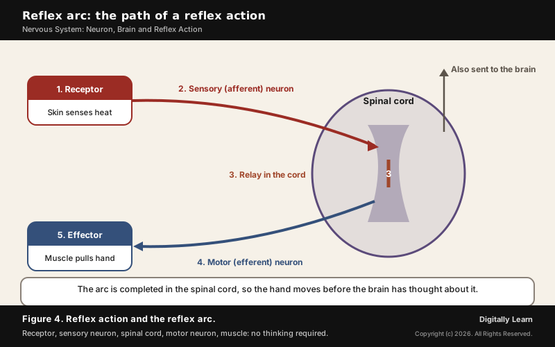
- Receptor: Nerve endings in the skin detect the heat.
- Sensory neuron: Carries the impulse to the spinal cord.
- Spinal cord: Relays the impulse to a motor neuron; nerves from all over the body meet here on their way to the brain, so the arc is completed in the cord itself.
- Motor neuron: Carries the order to the muscle.
- Effector: The muscle contracts and the hand moves away.
Reflex actions are therefore controlled by the spinal cord, not the brain, although the information also travels on to the brain. Reflex arcs evolved because thinking is not fast enough for danger, and even in animals with complex networks of neurons they remain more efficient for quick responses.
Parts of Brain and Their Functions
Forebrain: Cerebrum, Thalamus, Hypothalamus and Limbic System
The human brain has three major parts: the forebrain, the midbrain and the hindbrain. The forebrain is the main thinking part and is made of the cerebrum, the thalamus and the hypothalamus.
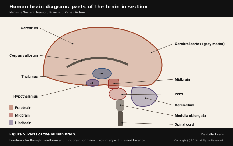
The cerebrum forms the major part of the human brain. A deep cleft splits it into left and right cerebral hemispheres, joined by a band of nerve fibres called the corpus callosum. Its folded outer layer, the cerebral cortex, is called grey matter because the neuron cell bodies gathered there give it a greyish colour; the myelinated fibres beneath form the white matter. The cortex has motor areas, sensory areas and association areas, which handle complex work such as memory and communication; the cerebrum controls voluntary actions.
- Thalamus: A major coordinating centre for sensory and motor signals, around which the cerebrum wraps.
- Hypothalamus: Lies at the base of the thalamus and controls body temperature and the urge to eat and drink; its neurosecretory cells make the hypothalamic hormones.
- Limbic system: The inner parts of the hemispheres with deep structures such as the amygdala and hippocampus; with the hypothalamus it regulates sexual behaviour, emotional reactions such as excitement, pleasure, rage and fear, and motivation.
Midbrain and Hindbrain: Pons, Cerebellum and Medulla Oblongata
The midbrain lies between the thalamus and hypothalamus above and the pons below. A canal called the cerebral aqueduct runs through it, and its back carries four round swellings, the corpora quadrigemina.
The hindbrain is made of the pons, the cerebellum and the medulla oblongata. The midbrain, pons and medulla together form the brain stem, which connects the brain to the spinal cord.
| Part | What it controls |
|---|---|
| Cerebrum (forebrain) | Thinking, voluntary actions, memory and the senses; it forms the major part of the brain |
| Hypothalamus (forebrain) | Body temperature, hunger and thirst; it also makes hormones |
| Pons (hindbrain) | Fibre tracts that link different regions of the brain |
| Cerebellum (hindbrain) | Precise voluntary movement, posture and balance |
| Medulla oblongata (hindbrain) | Breathing, heart reflexes, blood pressure, gut secretions and vomiting |
The cerebellum has a very folded surface that makes room for many more neurons, and it keeps voluntary movements precise and the body balanced, which is why walking in a straight line or riding a bicycle depends on it. The medulla contains centres for breathing, heart and blood-vessel reflexes and gastric secretion, and controls involuntary actions such as blood pressure, salivation and vomiting; the swallowing centre lies in the medulla and pons.
Disorders of the Nervous System and Neurotechnology
Alzheimer's Disease, Parkinson's Disease and Dementia
Dementia is a condition that affects memory, thinking and the ability to carry out daily activities, caused by diseases that damage the brain. In 2021, 57 million people were living with dementia, over 60 per cent of them in low- and middle-income countries, with nearly 10 million new cases every year. It is the seventh leading cause of death worldwide.
- Alzheimer’s disease: The most common form of dementia, which may account for 60 to 70 per cent of cases.
- Parkinson’s disease: A brain condition that causes problems with movement, mental health, sleep and pain. It involves the gradual loss of dopamine-producing neurons in a region of the brain called the substantia nigra. Its cause is unknown, but air pollution, pesticides and solvents may raise the risk, and levodopa/carbidopa is the most effective medicine.
Epilepsy, Stroke and How Drugs of Abuse Act on the Brain
Epilepsy is a chronic brain disease marked by repeated seizures, brief episodes of involuntary movement; it is diagnosed after two or more unprovoked seizures. About 50 million people have it, nearly 80 per cent of them in low- and middle-income countries, and up to 70 per cent could live seizure-free with proper diagnosis and treatment. A stroke happens when blood flow to part of the brain is reduced or blocked and brain cells die; it is ischaemic when caused by a lack of blood flow and haemorrhagic when caused by bleeding.
Many drugs of abuse act directly on the nervous system; the commonly abused ones are opioids, cannabinoids and coca alkaloids.
- Opioids: Bind to opioid receptors in the central nervous system and the gut. Heroin (smack) is made from morphine, taken from the latex of the poppy, and it is a depressant that slows body functions.
- Cannabinoids: Act on cannabinoid receptors found mainly in the brain; marijuana, hashish, charas and ganja come from the Cannabis plant.
- Cocaine: From the coca plant; it interferes with the transport of the neurotransmitter dopamine and strongly stimulates the central nervous system.
India's response is led by the Ministry of Social Justice and Empowerment, the nodal ministry for drug demand reduction under the National Action Plan for Drug Demand Reduction. Its Nasha Mukt Bharat Abhiyaan began on 15 August 2020 in 272 of the most vulnerable districts and was extended to every district from 15 August 2023; the helpline 14446 is linked to the narcotics helpline MANAS (1933) and the Tele-MANAS mental health service.
Care for disorders of the mind runs through the National Mental Health Programme (NMHP), in place since 1982. Its District Mental Health Programme, added in 1996 on the Bellary model of Karnataka, has now been approved for 767 districts, and Tele-MANAS, announced in the Union Budget 2022, adds a round-the-clock tele-mental-health service as the programme's digital arm.
Brain-Computer Interfaces: How Neurotechnology Reads Nerve Signals
A brain-computer interface (BCI) is a system that measures brain activity and turns it into useful output, most commonly to work a computer or a robotic limb. It works because the brain's activity, the impulses described above, can be picked up electrically.
- Non-invasive: Electrodes or scanners outside the head, as in EEG, MEG and MRI.
- Partially invasive: Electrodes on the surface of the brain or inside its blood vessels.
- Invasive: Microelectrode arrays placed in the brain tissue itself.
BCIs are used to research, map, assist, augment or repair sensory and motor functions; researchers are working on BCIs that could restore movement to paralysed limbs by electrically stimulating the muscles. Because the brain can adapt, it can learn to handle signals from an implant much like signals from its own senses and muscles.
Previous Year UPSC-CSE Questions
Previous Year UPSC-CSE Questions By the end you will be able to draft model answers for the following UPSC questions. Each question carries a collapsible framework showing how to approach it in the exam.
- UPSC Prelims 2007 Prelims-GSWhich one of the following parts of the human brain is the regulating centre for swallowing and vomiting?
How to approach this Prelims question
Approach: Ask whether swallowing and vomiting are voluntary or involuntary, then find the part that runs involuntary reflexes.
Trap to watch: The pons shares the swallowing centre, which makes it tempting, but vomiting is controlled by the medulla, so only the medulla covers both.
Key facts to recall:
- (a) Cerebellum: wrong. It handles balance and the precision of movement, not swallowing or vomiting.
- (b) Cerebrum: wrong. It controls voluntary actions, while swallowing and vomiting run as involuntary reflexes.
- (c) Medulla oblongata: right. It controls vomiting, salivation and blood pressure, and the swallowing centre lies in the medulla and pons.
- (d) Pons: wrong. It shares the swallowing centre, but vomiting is controlled by the medulla, so only the medulla covers both.
Answer signal: The medulla oblongata, so option (c) is the answer.
Sources and Further Reading
- NCERT: Biology, Class 11, Chapter 18, Neural Control and Coordination
- NCERT: Science, Class 10, Chapter 6, Control and Coordination
- NCERT: Biology, Class 11, Chapter 15, Body Fluids and Circulation
- NCERT: Biology, Class 12, Chapter 7, Human Health and Disease
- World Health Organization: Dementia, fact sheet
- World Health Organization: Parkinson disease, fact sheet
- World Health Organization: Epilepsy, fact sheet
- Press Information Bureau: Nasha Mukt Bharat Abhiyaan, 1 April 2026
- Department of Social Justice and Empowerment: Nasha Mukt Bharat Abhiyaan
- Directorate General of Health Services: National Mental Health Programme
- Wikipedia: Autonomic nervous system
- Wikipedia: Neurotransmitter
- Wikipedia: Cerebrospinal fluid
- Wikipedia: Medulla oblongata
- Wikipedia: Swallowing
- Wikipedia: Parkinson's disease
- Wikipedia: Stroke
- Wikipedia: Brain-computer interface
Editorial Disclaimer
This article is for UPSC preparation and explains how the human nervous system works. It is not medical advice for any individual.


