


Overview
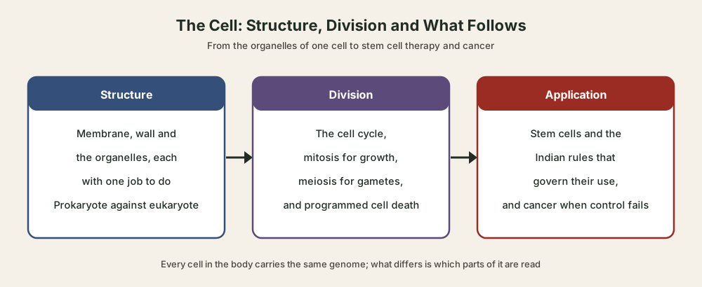
Cell structure is how a living cell is built, and cell function is what each part does. The cell is the basic structural and functional unit of all living organisms. It is either prokaryotic, with no membrane-bound nucleus, or eukaryotic, with one. The plasma membrane decides what enters, the organelles divide the work of life between them, and the gap between the two kinds of cell is what lets an antibiotic kill a bacterium and spare the patient.
Cell Theory and Types of Cells: Prokaryotic and Eukaryotic
Cell Theory: Schleiden, Schwann and Virchow
The cell is the fundamental structural and functional unit of all living organisms: anything less than a complete cell cannot live independently. Robert Hooke first observed a cell in 1665, in a thin slice of cork. Anton von Leeuwenhoek first saw and described a live cell, and Robert Brown later discovered the nucleus.
- Matthias Schleiden, 1838: A German botanist, he examined many plants and found that all plants are made of different kinds of cells, which form the plant’s tissues.
- Theodore Schwann, 1839: A German zoologist, he found that animal cells have a thin outer layer, today called the plasma membrane, and concluded that plants and animals are made of cells and products of cells.
- Rudolf Virchow, 1855: He explained that cells divide and new cells form from pre-existing cells, Omnis cellula-e cellula, which gave the theory its final shape.
Cell theory as understood today has two parts: all living organisms are composed of cells and products of cells, and all cells arise from pre-existing cells. Viruses sit outside it: all viruses live inside host cells.
Difference Between Prokaryotic and Eukaryotic Cell
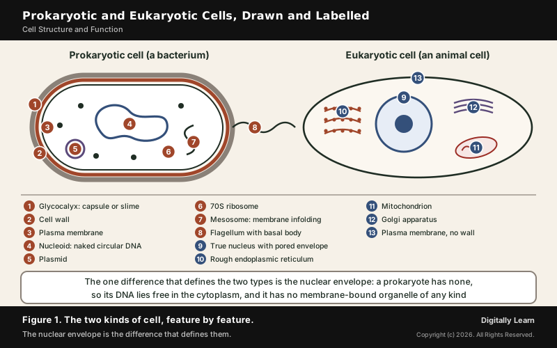
Cells with a membrane-bound nucleus are eukaryotic; cells without one are prokaryotic. Bacteria, blue-green algae, mycoplasma and PPLO (pleuro-pneumonia-like organisms) are prokaryotes. They are generally smaller and multiply more rapidly than eukaryotic cells. In both kinds, a semi-fluid matrix called the cytoplasm fills the cell and is the main arena of its chemical activity.
| Feature | Prokaryotic cell | Eukaryotic cell |
|---|---|---|
| Nucleus | No nuclear membrane; DNA lies naked in the cytoplasm | True nucleus bounded by a nuclear envelope |
| Genetic material | Single circular DNA, often with plasmids | Chromatin of DNA and protein inside the nucleus |
| Ribosomes | 70S (50S and 30S subunits) | 80S (60S and 40S subunits) |
| Membrane-bound organelles | Absent; a mesosome forms by infolding of the membrane | Present: ER, Golgi, lysosomes, mitochondria |
| Envelope | Glycocalyx, cell wall and plasma membrane | Cell wall in plants and fungi only |
| Examples | Bacteria, blue-green algae, mycoplasma, PPLO | Plants, animals, fungi, protists |
- Shapes of bacteria: Bacillus (rod-like), coccus (spherical), vibrio (comma-shaped) and spirillum (spiral).
- Gram stain: Bacteria that take up the Gram stain are Gram positive; those that do not are Gram negative.
- Surface structures: A bacterial flagellum has a filament, a hook and a basal body; pili and fimbriae do not help movement, but fimbriae help some bacteria attach to host tissues.
- Inclusion bodies: Reserve material such as phosphate, cyanophycean and glycogen granules lies free in the cytoplasm, not bound by any membrane.
Many bacteria carry small circular DNA outside the main chromosome, called plasmids. A plasmid gives the bacterium special characters, and one of them is resistance to antibiotics: the reason plasmids matter in the antibiotics section below.
Cell Size and Shape: Why Most Cells Stay Small
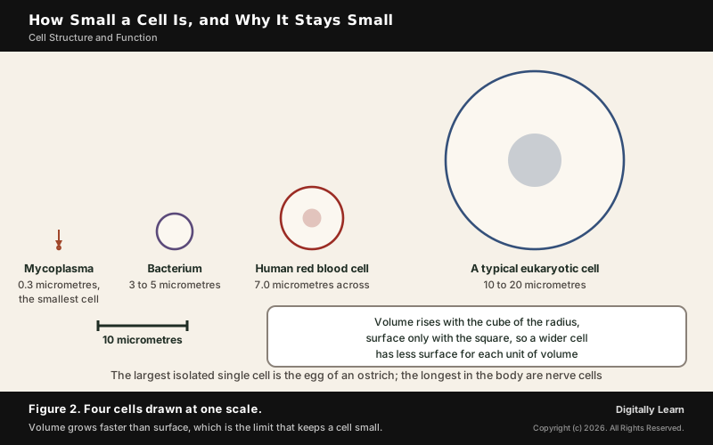
- Smallest: Mycoplasmas are only 0.3 micrometres long; bacteria are 3 to 5 micrometres.
- Largest: The largest isolated single cell is the egg of an ostrich.
- Human cells: A red blood cell is about 7.0 micrometres across; nerve cells are some of the longest cells.
- Shape follows function: Red blood cells are round and biconcave, columnar epithelial cells long and narrow, nerve cells branched and long, and tracheids elongated.
Size has a limit set by geometry. For a given shape, the surface area to volume ratio falls as the object grows: a cube 2 centimetres on a side has half the ratio of a cube 1 centimetre on a side. The larger this ratio, the more surface there is for each unit of volume through which material can diffuse, so a small cell exchanges food and waste with its surroundings far more easily than a large cell.
Plasma Membrane: Fluid Mosaic Model and Transport
Fluid Mosaic Model of the Plasma Membrane
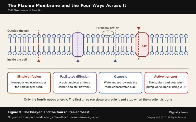
The plasma membrane is made mainly of lipids arranged in a bilayer, with the polar heads facing outwards and the non-polar tails inside, where they are protected from the watery surroundings. It also carries protein and carbohydrate. In the human red blood cell membrane, protein makes up about 52 per cent and lipid about 40 per cent.
- Peripheral proteins: Lie on the surface of the membrane.
- Integral proteins: Are partially or totally buried in it.
- Singer and Nicolson, 1972: Proposed the fluid mosaic model, now widely accepted.
The model's central idea is the quasi-fluid nature of the lipid, which lets proteins move sideways within the bilayer. This fluidity matters for cell growth, the formation of intercellular junctions, secretion, endocytosis and cell division.
Diffusion, Osmosis and Active Transport Across the Membrane
The membrane is selectively permeable: it lets some molecules through and not others. Transport across it takes two forms, and the difference is whether the cell spends energy.
- Passive transport: Neutral solutes move by simple diffusion along the concentration gradient, from higher to lower concentration, with no energy spent.
- Osmosis: The movement of water by diffusion.
- Carrier proteins: Polar molecules cannot pass the non-polar lipid bilayer, so a carrier protein of the membrane helps them across.
- Active transport: Some ions and molecules move against their concentration gradient, from lower to higher concentration. This uses ATP; the sodium-potassium pump is the standard example.
Plant roots show why the difference matters. Minerals in the soil are charged ions at a lower concentration than inside the root, so most cannot enter passively. Proteins in the membranes of root hair cells pump the ions in, using energy in the form of ATP. Mineral uptake is therefore a job of the plasma membrane.
Cell Wall in Plants, Fungi and Bacteria
Outside the membrane, plants and fungi carry a cell wall, a non-living rigid covering. It gives the cell its shape, protects it from mechanical damage and infection, helps cells interact with each other and keeps out unwanted large molecules.
- Plant cell walls: Cellulose, hemicellulose, pectins and proteins; in algae, cellulose, galactans, mannans and minerals like calcium carbonate.
- Bacteria: A cell envelope of three layers, the glycocalyx outside, then the cell wall, then the plasma membrane. Peptidoglycan, a mesh of sugars and amino acids, surrounds the bacterial membrane.
- Animal cells: No wall at all; the plasma membrane is the outer boundary.
A young plant cell has a primary wall that can grow; as the cell matures, a secondary wall forms on its inner side. The middle lamella, a layer mainly of calcium pectate, glues neighbouring cells together, and both wall and lamella are crossed by plasmodesmata, which connect the cytoplasm of neighbouring cells.
Cell Organelles and Their Functions
Nucleus, Chromatin and Nucleolus
The nucleus is enclosed by a nuclear envelope of two parallel membranes with a space of 10 to 50 nanometres between them, the perinuclear space. The outer membrane usually stays continuous with the endoplasmic reticulum and bears ribosomes. Minute nuclear pores let RNA and protein molecules pass both ways between the nucleus and the cytoplasm.
- Chromatin: Extended nucleoprotein fibres present when the nucleus is not dividing; they condense into chromosomes when the cell divides.
- Nucleolus: A spherical body inside the nucleus that is not bound by a membrane; it is the site of active ribosomal RNA synthesis.
- Centromere: Holds the two chromatids of a chromosome; its position gives four types: metacentric, sub-metacentric, acrocentric and telocentric.
A cell normally has one nucleus, but the number varies, and some mature cells have none at all: the red blood cells of many mammals and the sieve tube cells of vascular plants.
A cell that is actively making protein has larger and more numerous nucleoli. How chromosomes are copied and shared out when the cell divides is the subject of the article on the cell cycle and cell division.
Ribosomes, Endoplasmic Reticulum, Golgi Apparatus and Lysosomes
Protein is made on ribosomes, first seen under the electron microscope as dense particles by George Palade in 1953. They are made of RNA and protein and are not surrounded by any membrane, and they carry out this work in every cell, prokaryotic and eukaryotic.
- Two subunits: The 80S ribosome of eukaryotes has 60S and 40S subunits; the 70S ribosome of prokaryotes has 50S and 30S.
- S for Svedberg: The unit is a sedimentation coefficient, an indirect measure of density and size, so subunit values do not simply add up.
- Polysome: Several ribosomes may attach to one messenger RNA and translate it together.
- Where found: In the cytoplasm, on the rough ER, and inside chloroplasts and mitochondria.
- Rough ER: Endoplasmic reticulum with ribosomes on its surface; common in cells making protein for secretion.
- Smooth ER: No ribosomes; the main site of lipid synthesis, and of steroidal hormones in animal cells.
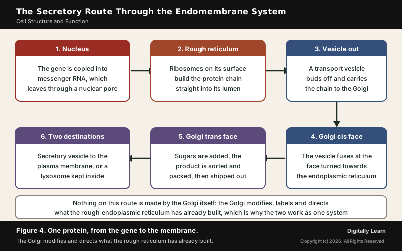
- Golgi apparatus: Stacks of flat cisternae with a cis (forming) face and a trans (maturing) face. Materials from the ER are modified here, and it is the main site of glycoprotein and glycolipid formation.
- Microbodies: Minute membrane-bound vesicles containing various enzymes, found in both plant and animal cells.
- Lysosomes: Membrane-bound vesicles packed in the Golgi, rich in hydrolytic enzymes (lipases, proteases, carbohydrases) that work best at acidic pH.
These organelles work as one endomembrane system. A protein for export is built on the rough ER, carried to the cis face of the Golgi, modified, and released from the trans face towards the membrane or a lysosome. Because their enzymes can digest carbohydrates, proteins, lipids and nucleic acids, lysosomes break down worn-out material inside the cell.
Mitochondria and Chloroplasts: Organelles With Their Own DNA
The sites of aerobic respiration are the mitochondria. They produce cellular energy as ATP, which is why they are called the power houses of the cell. Each is usually sausage-shaped, 0.2 to 1.0 micrometres across, with a double membrane: the inner one folds into cristae, which increase the surface area.
In green plants, most chloroplasts lie in the mesophyll cells of the leaves. They are 5 to 10 micrometres long and 2 to 4 micrometres wide, and like mitochondria they have a double membrane. Inside, the stroma holds flattened sacs called thylakoids, stacked like piles of coins into grana. Chlorophyll traps the light energy that photosynthesis needs.
| Feature | Mitochondrion | Chloroplast |
|---|---|---|
| Job | Aerobic respiration, ATP | Photosynthesis |
| Inner folds | Cristae | Thylakoids stacked into grana |
| Own genetic material | Single circular DNA, a few RNAs, 70S ribosomes | Small double-stranded circular DNA, 70S ribosomes |
Circular DNA and 70S ribosomes, features they share with bacteria, fit the endosymbiotic theory: mitochondria and chloroplasts descend from free-living prokaryotes taken inside another cell. Mitochondria also divide by fission. In most animals, mitochondrial DNA is inherited from the mother.
Cytoskeleton, Centrioles, Cilia and Flagella
The cytoskeleton is a network of protein filaments, the microtubules, microfilaments and intermediate filaments. It gives mechanical support, holds the cell's shape and supports movement.
- Cilia and flagella: Hair-like outgrowths of the membrane; cilia are small, flagella longer. Both have an axoneme of nine doublets of microtubules around a central pair, the 9+2 array, joined by nine radial spokes.
- Basal body: Both grow from a centriole-like structure called the basal body.
- Centrosome: Two centrioles at right angles, each with nine evenly spaced triplet fibrils of tubulin, arranged like a cartwheel. Centrioles form the basal body of cilia and flagella and the spindle fibres of cell division.
- Plant cells: Centrioles are absent in conifers and flowering plants.
Cilia at work can be seen in the body. Ciliated epithelium moves particles or mucus in one direction over its surface, and lines hollow organs such as the bronchioles and the fallopian tubes.
Difference Between Plant Cell and Animal Cell
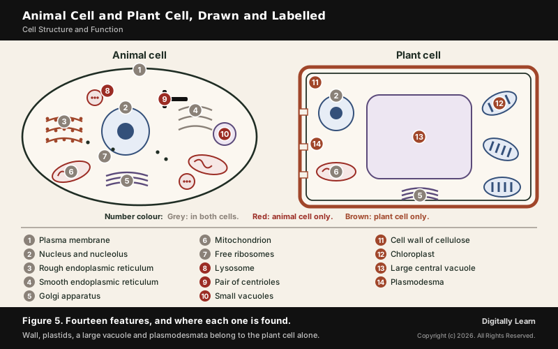
Plant Cell and Animal Cell Diagram and Comparison Table
Plant and animal cells share most organelles. The difference between plant cell and animal cell comes down to a short list, and one item, the way the cell divides, follows directly from the rigid wall of the plant cell.
| Feature | Plant cell | Animal cell |
|---|---|---|
| Cell wall | Present, of cellulose | Absent |
| Plastids | Present, including chloroplasts | Absent |
| Vacuole | Large central vacuole, up to 90 per cent of the volume | Sometimes present, never as large |
| Centrioles | Absent in flowering plants and conifers | Present in the centrosome |
| Stored carbohydrate | Starch | Glycogen |
| Cell division (cytokinesis) | Cell plate forms in the centre | Furrow in the membrane deepens inwards |
Because the wall does not stretch, a dividing plant cell cannot pinch itself in two. It builds a cell plate in the centre that grows outwards, while an animal cell forms a furrow that deepens until the cytoplasm splits.
Plastids, Vacuoles and Connections Between Cells
- Chloroplast pigments: Hold chlorophyll and carotenoids, which trap light for photosynthesis.
- Chromoplasts: Hold fat-soluble carotenoids that give parts of the plant a yellow, orange or red colour.
- Leucoplasts: Colourless stores: amyloplasts store starch (as in potato), elaioplasts oils and fats, and aleuroplasts proteins.
The vacuole is bound by a single membrane, the tonoplast, and holds water, sap, excretory products and other materials. In plant cells it can take up to 90 per cent of the cell's volume. The tonoplast moves ions into the vacuole against their concentration gradient, so they are far more concentrated there than in the cytoplasm.
- Contractile vacuole: In Amoeba, it handles osmoregulation and excretion.
- Food vacuoles: Many protists form them by engulfing food particles.
- Plasmodesmata: Connect the cytoplasm of neighbouring plant cells through the wall.
Biomolecules and Enzymes in the Cell
Carbohydrates, Proteins, Lipids and Nucleic Acids
- Carbohydrates: Polysaccharides are long chains of sugars. Cellulose is a polymer of glucose alone; starch stores energy in plants and glycogen in animals.
- Proteins: Chains of amino acids linked by peptide bonds, drawn from 20 types of amino acid. Collagen is the most abundant protein in the animal world, and RuBisCO the most abundant in the whole biosphere.
- Lipids: Fatty acids esterified with glycerol form monoglycerides, diglycerides and triglycerides; phospholipids, such as lecithin, are found in the cell membrane.
- Nucleic acids: Built from nitrogen bases (adenine, guanine, cytosine, uracil and thymine) joined to a sugar and a phosphate, forming nucleotides.
- Primary structure: The sequence of amino acids, from the N-terminal to the C-terminal end.
- Secondary structure: Parts of the chain fold into a helix, like a revolving staircase, or a pleated sheet.
- Tertiary structure: The whole chain folds on itself like a hollow woollen ball, the 3-dimensional shape most biological activity needs.
- Quaternary structure: The arrangement of several chains; adult human haemoglobin has four subunits, two alpha and two beta.
A simple way to hold these together is by job: carbohydrates and lipids are mainly fuel and structure, proteins are the cell's machinery and structure, and nucleic acids carry information. Enzymes, the machinery that runs every reaction, are the subject of the next section.
Enzymes: Active Site, Activation Energy and Inhibition
An enzyme is a protein that catalyses a reaction in the cell. It lowers the activation energy, the energy barrier a substrate must cross to become a product, and comes out of the reaction unchanged.
- Active site: A crevice or pocket in the folded enzyme into which the substrate fits; this fit makes the enzyme specific.
- Optimum conditions: Each enzyme is most active at an optimum temperature and pH; low temperature keeps it temporarily inactive, while high temperature destroys it.
- Cofactors: Non-protein helpers: prosthetic groups, co-enzymes and metal ions.
- Ribozymes: Almost all enzymes are proteins, but some nucleic acids also act as enzymes.
- Competitive inhibition: An inhibitor that closely resembles the substrate competes for the active site; malonate inhibits succinic dehydrogenase this way. Such inhibitors are used against bacterial pathogens.
| Class | What the enzymes do |
|---|---|
| Oxidoreductases (dehydrogenases) | Catalyse oxidation and reduction between two substrates |
| Transferases | Transfer a group other than hydrogen from one substrate to another |
| Hydrolases | Break bonds by hydrolysis, such as ester, peptide and glycosidic bonds |
| Lyases | Remove groups by other means, leaving double bonds |
| Isomerases | Convert one isomer into another |
| Ligases | Join two compounds together |
Antibiotics and Antimicrobial Resistance
How Antibiotics Target the Bacterial Cell
An antibiotic kills bacteria or stops them growing without harming the patient. It can do this because it blocks a life process that bacteria have and human cells do not.
- Bacterial cell wall: Many bacteria build a wall. Penicillin blocks the processes that build it, so growing bacteria die easily, and human cells make no wall to damage. It works by interfering with the making of peptidoglycan.
- Bacterial ribosomes: Protein synthesis inhibitors exploit the major differences between prokaryotic and eukaryotic ribosomes, binding subunits such as the 50S of the bacterial 70S ribosome.
- Folate: Sulfonamides block an enzyme that bacteria use to make folate. Humans take folate (vitamin B9) from the diet instead.
The same logic shows what antibiotics cannot do. Viruses do not use these pathways at all, so antibiotics do not work against viral infections: taking them for a common cold does not shorten or ease it. They help only against a bacterial infection that comes along with the virus.
How Antimicrobial Resistance Develops and Spreads
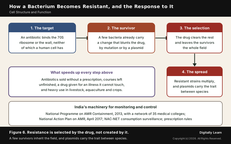
Bacteria become resistant through spontaneous mutation, horizontal gene transfer and the selective pressure of antibiotic overuse, in medicine and in agriculture. Heavy use of a drug is a form of natural selection: the bacteria that survive it multiply, so resistant organisms appear within months or years rather than centuries. It is evolution driven by human action.
- Down the generations: A resistant bacterium divides into resistant daughters.
- Between bacteria: Genes for antibiotic resistance can pass from one species of bacteria to another; this horizontal gene transfer, often involving plasmids, is the main way resistance spreads.
- Drivers: Misuse and overuse of antibiotics, such as taking them when they are not needed or taking the wrong type, remain one of the main drivers; overprescribing and inappropriate prescription have accelerated the problem.
- Farms: Antimicrobials are used across livestock, aquaculture and crop production, so resistance also grows outside hospitals.
The scale is large. Bacterial resistance was associated with more than 4.7 million deaths worldwide in 2021, and about one in six laboratory-confirmed bacterial infections in 2023 were resistant to antibiotics.
India's Response to Antimicrobial Resistance
- National Programme on AMR Containment, 2013: Coordinated by the National Centre for Disease Control, the nodal agency; it supports a network of 35 state medical college laboratories that report resistance data.
- National Action Plan on AMR, April 2017: One of its strategic priorities is to optimise the use of antimicrobials; infection prevention and control is the third.
- Infection control guidelines: National guidelines for healthcare facilities were released in January 2020.
- Consumption surveillance: The National Antimicrobial Consumption Network collects data from 35 tertiary care institutions. Over 2017 to 2021, cephalosporins were the most consumed class, and the Reserve group of antibiotics ranged from 1 to 5 per cent of use.
- Regulation of drugs: Medicines are regulated under the Drugs and Cosmetics Act, 1940 and the Rules of 1945.
No single sector can solve the problem alone, because resistant bacteria move between people, animals and the environment. The World Health Organization's global action plan, carried out through national plans such as India's, therefore follows a One Health approach, treating human, animal and environmental health together.
Previous Year UPSC-CSE Questions
Previous Year UPSC-CSE Questions By the end you will be able to draft model answers for the following UPSC questions. Each question carries a collapsible framework showing how to approach it in the exam.
- UPSC Prelims 1996 Prelims-GSMatch List I (Physiological processes) with List II (Cell organelles), where List II reads A) Plasma membrane, B) Chloroplast, C) Mitochondria, D) Ribosomes:
- I. Photosynthesis
- II. Mineral uptake
- III. Respiration
- IV. Protein Synthesis
Select the correct answer by using the codes given below the lists:
How to approach this Prelims question
Approach: Place the two obvious pairs first, photosynthesis with the chloroplast and protein synthesis with the ribosome, then decide between the plasma membrane and the mitochondrion for the remaining two.
Trap to watch: Mineral uptake is a transport event at the boundary of the cell, so it belongs to the plasma membrane, not to an organelle.
Key facts to recall:
- (a) I-A, II-B, III-C, IV-D: wrong. Photosynthesis is not done at the plasma membrane, and mineral uptake is not done in the chloroplast.
- (b) I-A, II-B, III-D, IV-C: wrong. It swaps the first two pairs and also the last two.
- (c) I-B, II-A, III-C, IV-D: right. Chloroplast for photosynthesis, plasma membrane for mineral uptake, mitochondria for respiration, ribosomes for protein synthesis.
- (d) I-B, II-A, III-D, IV-C: wrong. Respiration happens in the mitochondria and protein synthesis on the ribosomes, not the other way round.
Answer signal: Photosynthesis with B, mineral uptake with A, respiration with C and protein synthesis with D, so option (c) is the answer.
- UPSC Prelims 2001 Prelims-GSWhich of the following cell organelles play the most significant role in protein synthesis?
How to approach this Prelims question
Approach: Identify the organelle that builds the peptide chain, then the organelle whose surface carries it when the product is destined for export.
Trap to watch: The Golgi apparatus modifies and packages protein but does not build it, so a pair naming the Golgi is incorrect.
Key facts to recall:
- (a) Lysosome and Centrosome: wrong. The lysosome digests, and the centrosome organises the spindle.
- (b) Endoplasmic reticulum and Ribosome: right. Ribosomes make the protein, and the rough endoplasmic reticulum is studded with them.
- (c) Golgi apparatus and Mitochondria: wrong. The Golgi packages proteins after they are made, and mitochondria supply energy.
- (d) Lysosome and Mitochondria: wrong. Neither is a site of protein synthesis.
Answer signal: The endoplasmic reticulum and the ribosome, so option (b) is the answer.
- UPSC Prelims 2001 Prelims-GSWhich organelle in the cell, other than nucleus, contains DNA?
How to approach this Prelims question
Approach: Recall which organelles are semi-autonomous, that is, which divide by themselves and carry their own genetic apparatus.
Trap to watch: The chloroplast also carries DNA but is absent from animal cells and is not among the options here.
Key facts to recall:
- (a) Centriole: wrong. Centrioles organise the spindle and carry no DNA of their own.
- (b) Golgi apparatus: wrong. It packages and dispatches proteins and holds no DNA.
- (c) Lysosome: wrong. It is a sac of digestive enzymes without DNA.
- (d) Mitochondrion: right. Its matrix holds circular DNA and 70S ribosomes.
Answer signal: The mitochondrion, so option (d) is the answer.
- UPSC Mains 2014 GS-IIICan overuse and free availability of antibiotics without Doctor’s prescription, be contributors to the emergence of drug-resistant diseases in India? What are the available mechanisms for monitoring and control? Critically discuss the various issues involved.
Answer outline: how to structure the answer
What the question asks, the main arguments, relevant examples and the direction of the conclusion. This is not a full model answer.
Introduction: Resistance is selected rather than created: an antibiotic clears the susceptible majority of a bacterial population and leaves the resistant few to multiply, so the more often a drug is used, and the less completely, the faster resistance spreads.
Body (sub-themes to develop):
- Yes to the premise: selection pressure, vertical spread by division and horizontal spread by plasmids between species; unfinished courses and antibiotics taken for viral illness add pressure without benefit.
- Scale: the World Health Organization associates more than 4.7 million deaths worldwide with bacterial resistance in 2021, and roughly one in six laboratory-confirmed bacterial infections in 2023 did not respond.
- Mechanisms for monitoring: the National Programme on AMR Containment of 2013 with its network of 35 medical colleges, and the National Antimicrobial Consumption Network, whose 2017 to 2021 report found cephalosporins the most consumed class.
- Mechanisms for control: the National Action Plan of April 2017, infection prevention and control as its third strategic priority with national guidelines issued in January 2020, and prescription control under the Drugs and Cosmetics Act administered by the drugs regulator.
- Critical issues: enforcement of prescription rules rests largely with State drug controllers, surveillance covers tertiary hospitals rather than the primary care where most prescribing happens, and animal and crop use sits outside health administration, which is why the plan is framed as One Health.
Conclusion: Free availability does contribute, and India has built the surveillance and the legal instruments to answer it; what remains unfinished is enforcement at the counter and coverage beyond tertiary hospitals and beyond human medicine.
Sources and Further Reading
- NCERT: Biology, Class 11, Chapter 8, Cell: The Unit of Life
- NCERT: Biology, Class 11, Chapter 9, Biomolecules
- NCERT: Science, Class 9, Chapter 2, Cell: The Building Block of Life
- NCERT: Biology, Class 12, Chapter 10, Biotechnology and its Applications
- World Health Organization: Antimicrobial resistance, fact sheet
- National Centre for Disease Control: Antimicrobial Resistance (AMR) Containment
- National Centre for Disease Control: Surveillance of Antimicrobial Consumption under NAC-NET, July 2023
- Food and Agriculture Organization of the United Nations: Antimicrobial Resistance
- Central Drugs Standard Control Organisation: Acts and Rules, including the Drugs and Cosmetics Act 1940 and Rules 1945
- Wikipedia: Peptidoglycan
- Wikipedia: Cell junction
- Wikipedia: Fluid mosaic model
Editorial Disclaimer
This article is for UPSC preparation and explains how a cell is built and how its parts work. Points on medicines and their regulation follow the official documents cited, which govern any question of practice.


