

Overview
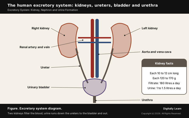
The excretory system removes the nitrogenous wastes and excess water and salts that the body produces. In humans it is made of two kidneys, two ureters, a urinary bladder and a urethra, and each kidney does its work through about a million tiny filtering units called nephrons.
Excretion and the Three Kinds of Nitrogenous Waste
Ammonotelic, Ureotelic and Uricotelic Animals
The work of every excretory system begins with the wastes an animal makes. Animals build up ammonia, urea, uric acid, carbon dioxide, water and ions such as sodium, potassium and chloride, from their metabolism or from taking in too much, and these must be removed wholly or in part. The most important are the three nitrogenous wastes, and animals are grouped by which one they mainly excrete.
| Waste | Animals that excrete it | Water needed |
|---|---|---|
| Ammonia, the most toxic | Ammonotelic: many bony fishes, aquatic amphibians, aquatic insects | A large amount |
| Urea, less toxic | Ureotelic: mammals, many land amphibians, marine fishes | Moderate |
| Uric acid, the least toxic | Uricotelic: reptiles, birds, land snails, insects | Very little |
Water decides the choice. Ammonia dissolves readily, so aquatic animals simply let it diffuse out across the body surface or the gills. Life on land called for conserving water, which is why land animals make less toxic urea or uric acid instead. In mammals the liver converts ammonia into urea, the blood carries it to the kidneys, and the kidneys filter it out; birds and reptiles pass uric acid as a pellet or paste with hardly any water lost.
Excretory Organs in Animals: Flame Cells to Kidneys
Excretory organs range from simple tubes in invertebrates to the complex kidneys of vertebrates. Besides removing nitrogenous waste, most of them also keep the balance of water and ions, which is called osmoregulation.
- Protonephridia or flame cells: Flatworms such as Planaria, rotifers, some annelids and the cephalochordate Amphioxus; they mainly regulate fluid and ions.
- Nephridia: Earthworms and other annelids; they remove nitrogenous waste and balance fluid and ions.
- Malpighian tubules: Most insects, including cockroaches.
- Antennal or green glands: Crustaceans such as prawns.
- Kidneys: All vertebrates, including humans.
Excretory System in Human Beings: Kidneys, Ureters, Bladder and Urethra
Human Kidney Diagram: Position and Structure of the Kidney
The human excretory system is made of a pair of kidneys, a pair of ureters, a urinary bladder and a urethra. The kidneys are located at the back of the abdomen, one on either side of the backbone, between the level of the last thoracic and the third lumbar vertebra. Urine made in the kidneys runs down the ureters to the urinary bladder, where it is stored until it leaves through the urethra.
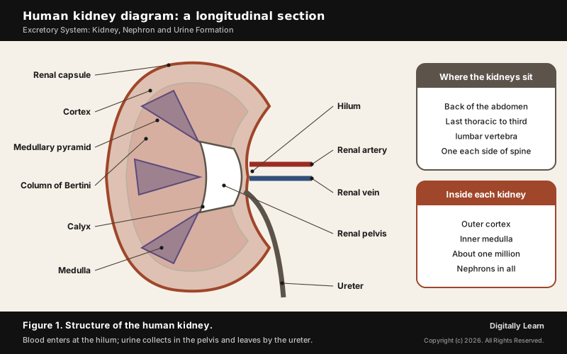
- Size: Each adult kidney is a reddish-brown, bean-shaped organ 10 to 12 cm long, 5 to 7 cm wide and 2 to 3 cm thick, weighing 120 to 170 g.
- Hilum: A notch on the inner, concave side where the renal artery enters and the renal vein and the ureter leave.
- Renal pelvis and calyces: A funnel-shaped space inside the hilum, with projections called calyces that collect urine.
- Cortex and medulla: Under a tough outer capsule lie an outer cortex and an inner medulla; the medulla forms cone-shaped medullary pyramids, and the cortex runs between them as the columns of Bertini.
Nephron Diagram and Structure: Glomerulus, Bowman's Capsule and Tubules
A nephron is the functional unit of the kidney, the tiny tube in which urine is made, and each kidney holds nearly one million of them. Every nephron has two parts: the glomerulus, a tuft of capillaries, and the renal tubule.
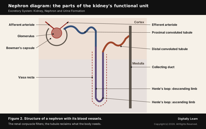
- Glomerulus: A tuft of capillaries formed by the afferent arteriole, a fine branch of the renal artery; blood leaves it by the efferent arteriole.
- Bowman’s capsule: A double-walled cup that encloses the glomerulus; the two together form the Malpighian body or renal corpuscle.
- Proximal convoluted tubule (PCT): A highly coiled part that follows the capsule.
- Loop of Henle: A hairpin loop with a descending and an ascending limb.
- Distal convoluted tubule (DCT): A second coiled part; the DCTs of many nephrons open into a collecting duct, and collecting ducts empty into the renal pelvis.
The renal corpuscle, PCT and DCT lie in the cortex, while the loop of Henle dips into the medulla. In most nephrons the loop is short, and these are cortical nephrons; in some it runs deep into the medulla, and these are juxtamedullary nephrons. The efferent arteriole forms a network of peritubular capillaries around the tubule, and one small vessel of it runs beside the loop as the U-shaped vasa recta, which is absent or much reduced in cortical nephrons.
Urine Formation in the Nephron
Glomerular Filtration and the Glomerular Filtration Rate
Urine formation involves three processes, glomerular filtration, reabsorption and secretion, carried out in different parts of the nephron. The first step, filtration, happens in the glomerulus.
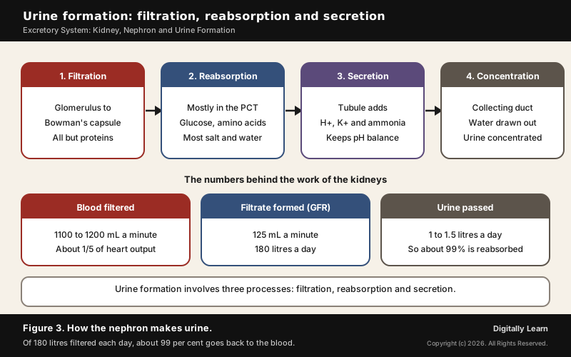
The kidneys filter on average 1100 to 1200 millilitres of blood a minute, roughly one-fifth of what each ventricle of the heart pumps out. The blood pressure in the glomerular capillaries pushes fluid through three layers: the capillary wall, the wall of Bowman's capsule, and a basement membrane between them. The capsule cells, called podocytes, leave minute filtration slits, so almost everything in plasma except the proteins passes through; that is why it is called ultrafiltration.
The volume of filtrate formed each minute is the glomerular filtration rate (GFR): about 125 millilitres a minute, or 180 litres a day, in a healthy person. The kidney keeps it steady through the juxtaglomerular apparatus (JGA), a sensitive region where the distal tubule touches the afferent arteriole. When GFR falls, the JG cells release renin, which raises the blood flow through the glomerulus and brings GFR back to normal.
Tubular Reabsorption and Secretion
Of the 180 litres of filtrate formed each day only about 1.5 litres leave as urine, so nearly 99 per cent is taken back into the blood; this is reabsorption. Glucose, amino acids and sodium are reabsorbed actively, nitrogenous wastes passively, and water passively in the first parts of the tubule. At the same time the tubule cells secrete hydrogen ions, potassium ions and ammonia into the filtrate, which keeps the ionic and acid-base balance of the body fluids.
| Segment | Main work | Detail |
|---|---|---|
| Proximal tubule (PCT) | Most reabsorption | Nearly all nutrients and 70 to 80 per cent of salts and water |
| Descending limb of Henle | Concentrates the filtrate | Lets water out; almost blocks salts |
| Ascending limb of Henle | Dilutes the filtrate | Keeps water in; moves salts out |
| Distal tubule (DCT) | Conditional reabsorption | Sodium and water as needed; keeps pH |
| Collecting duct | Final water recovery | Large amounts of water drawn back for concentrated urine |
Counter-Current Mechanism: How the Kidney Concentrates Urine
Mammals can produce a concentrated urine, and the loop of Henle and the vasa recta are what make this possible. Filtrate flows down one limb of the loop and up the other, in opposite directions, and blood flows the same way in the two limbs of the vasa recta. Such opposite flow is called a counter current.
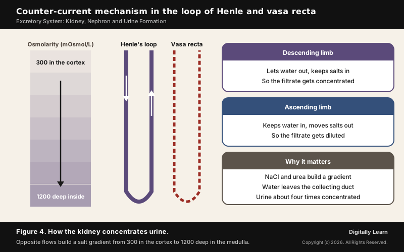
Because the two tubes run close together with opposite flows, they keep the fluid around them growing more concentrated towards the inner medulla, from about 300 mOsmol per litre in the cortex to about 1200 deep inside. Sodium chloride and urea build this gradient: the ascending limb of Henle's loop passes salt out, the vasa recta carries it back, and the collecting tubule returns some urea. Water then leaves the collecting duct easily, and human kidneys can make urine nearly four times as concentrated as the first filtrate.
Regulation of Kidney Function
ADH, the Renin-Angiotensin Mechanism and Atrial Natriuretic Factor
The kidneys are regulated by hormonal feedback involving the hypothalamus, the juxtaglomerular apparatus and, to some extent, the heart. The three mechanisms act on body water, salt and blood pressure.
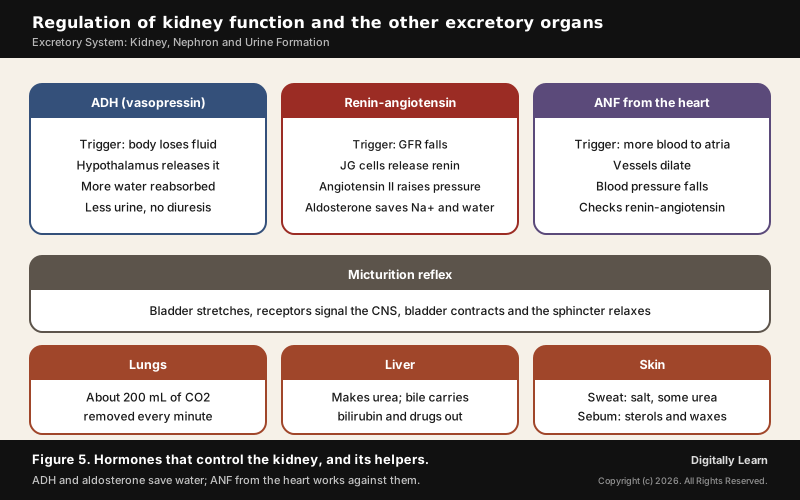
- ADH or vasopressin: When the body loses too much fluid, osmoreceptors stimulate the hypothalamus to release antidiuretic hormone from the neurohypophysis. ADH increases water reabsorption in the later parts of the tubule and so prevents heavy urine flow (diuresis); when fluid volume rises, its release is switched off.
- Renin-angiotensin mechanism: A fall in glomerular blood flow, pressure or GFR makes the JG cells release renin, which converts angiotensinogen to angiotensin I and then angiotensin II. Angiotensin II narrows blood vessels, raising glomerular pressure and GFR, and makes the adrenal cortex release aldosterone, which increases the reabsorption of sodium and water.
- Atrial natriuretic factor (ANF): More blood reaching the atria of the heart releases ANF, which widens blood vessels and lowers blood pressure, acting as a check on the renin-angiotensin mechanism.
Several of these hormones act through blood pressure; blood pressure itself, and why high pressure damages the kidneys, is explained in the circulatory system.
Micturition and the Excretory Role of Lungs, Liver and Skin
Micturition is the release of urine. As the bladder fills and stretches, receptors in its wall signal the central nervous system, which makes the bladder muscle contract and the urethral sphincter relax; this is the micturition reflex. An adult passes 1 to 1.5 litres of light-yellow, slightly acidic urine (pH about 6.0) a day, carrying 25 to 30 grams of urea.
Urine tests help diagnose disease: glucose in urine (glycosuria) and ketone bodies (ketonuria) point to diabetes mellitus. The kidneys are also not the only organs of excretion.
- Lungs: Remove about 200 millilitres of carbon dioxide a minute and a good deal of water every day; breathing is explained in the respiratory system.
- Liver: Converts ammonia into urea and secretes bile carrying bilirubin, biliverdin, cholesterol, used hormones, vitamins and drugs, which leave with the digestive wastes.
- Skin: Sweat carries sodium chloride, a little urea and lactic acid; sebum from the sebaceous glands carries sterols, hydrocarbons and waxes. Small amounts of nitrogenous waste also leave in saliva.
Kidney Disorders, Dialysis and Transplantation in India
Kidney Stones, Uraemia, Chronic Kidney Disease and Glomerulonephritis
When the kidneys fail, urea builds up in the blood, a condition called uraemia, which is highly harmful and can lead to kidney failure.
Chronic kidney disease (CKD) is a gradual loss of kidney function over time. Surveys in India put its prevalence at 8 to 17 per cent of the population, and about 10 to 20 per cent of people with CKD go on to end-stage kidney failure, the stage at which survival is not possible without dialysis or a kidney transplant. Chronic kidney disease is one of the conditions covered by India's National Programme for Prevention and Control of Non-Communicable Diseases. Two other disorders of the kidney are named separately.
- Renal calculi (kidney stones): Stones or insoluble masses of crystallised salts, such as oxalates, formed within the kidney.
- Glomerulonephritis: Inflammation of the glomeruli of the kidney.
Kidney stones form when urine carries more of a crystal-forming substance than it can hold dissolved and there is not enough fluid: tiny crystals appear and clump into hard masses in the upper urinary tract. Most stones contain calcium; calcium stones make up about 80 per cent of cases in the United States and usually contain calcium oxalate, alone or with calcium phosphate.
Haemodialysis and the Pradhan Mantri National Dialysis Programme
Dialysis is required when the kidneys can no longer clear urea from the blood. In haemodialysis, an artificial kidney does the filtering outside the body.
- Blood out: Blood is drawn from a convenient artery and an anticoagulant such as heparin is added.
- Through the dialyser: The blood passes through a coiled cellophane tube bathed in dialysing fluid that is like plasma but has no nitrogenous wastes, so urea and other wastes move out across the porous membrane.
- Blood back: Anti-heparin is added and the cleaned blood is pumped back into a vein.
Peritoneal dialysis is the other form. It can be done at home, is cheaper, and needs little infrastructure or technical staff, so it widens access for patients far from a hospital.
An artificial kidney differs from a real one in an important way: there is no reabsorption, only removal of wastes by diffusion. In India about 2.2 lakh new patients with end-stage renal disease are added each year, creating demand for 3.4 crore dialysis sessions, and its high cost can be financially catastrophic for families.
- Launch: The Pradhan Mantri National Dialysis Programme was announced in the Union Budget 2016-17 and rolled out on 7 April 2016 under the National Health Mission, in public-private partnership mode at district hospitals.
- Who it serves: Dialysis is free for Below Poverty Line patients; the programme has haemodialysis and peritoneal dialysis components.
- Reach: By 31 December 2022 it ran in all 36 States and Union Territories, in 641 districts, at 1,350 centres with 8,871 machines, and 17.27 lakh people had used it.
- Portability: A national portal launched on 5 May 2022 registers dialysis patients with their ABHA health ID, so treatment can continue anywhere under One Nation-One Dialysis.
Kidney Transplant and Organ Donation: the 1994 Act and NOTTO
A kidney transplant is the ultimate treatment for kidney failure: a working kidney is taken from a donor, preferably a close relative, which lowers the chance of the recipient's immune system rejecting it. Organ transplants are needed when a person's own organ has failed from disease or injury, and common transplants include corneas, kidneys, heart, liver, pancreas, lungs, intestines and bone marrow.
The Transplantation of Human Organs Act, 1994 regulates the removal, storage and transplantation of organs and bans commercial dealing in them. It recognises brain stem death as legal death: after natural cardiac death only a few tissues such as cornea, bone and skin can be donated, but after brain stem death almost 37 organs and tissues can be.
- 2011 amendment: Renamed the law the Transplantation of Human Organs and Tissues Act (THOTA), added tissues, widened near relatives to include grandparents and grandchildren, and allowed swap donation between unmatched pairs; it came into force on 10 January 2014 in the first States and Union Territories.
- NOTTO: The National Organ and Tissue Transplant Organisation, the apex body at Safdarjung Hospital, New Delhi, under the Directorate General of Health Services, keeps the national registry and networks with regional (ROTTO) and state (SOTTO) organisations.
- Scale: India performed over 18,900 organ transplants in 2024, up from fewer than 5,000 in 2013, and ranks third in the world after the United States and China.
Previous Year UPSC-CSE Questions
Previous Year UPSC-CSE Questions By the end you will be able to draft model answers for the following UPSC questions. Each question carries a collapsible framework showing how to approach it in the exam.
- UPSC Prelims 2003 Prelims-GSWith reference to the work of human kidney, consider the following statements:
- After the waste is removed in the kidney, the cleaner blood is sent back through renal artery.
- From Bowman’s capsule, the filtered liquid passes through tiny tubes where much of the glucose is reabsorbed and sent back to the blood in the renal vein.
Which of these statements is/are correct?
How to approach this Prelims question
Approach: Check the direction of each vessel, then check where glucose goes after filtration.
Trap to watch: Statement 1 swaps the vessels: the renal artery brings blood in; cleaned blood leaves by the renal vein.
Key facts to recall:
- (a) Only 1: wrong. Statement 1 swaps the vessels: the renal artery brings blood in, and cleaned blood leaves by the renal vein.
- (b) Only 2: right. The filtrate passes from Bowman's capsule into the tubules, where glucose is reabsorbed actively and returned to the blood.
- (c) Both 1 and 2: wrong. It includes the false statement 1.
- (d) Neither 1 nor 2: wrong. Statement 2 is correct.
Answer signal: Only statement 2 is correct, so option (b) is the answer.
- UPSC Prelims 2000 Prelims-GSThe stones formed in human kidney consist mostly of
How to approach this Prelims question
Approach: Pick the compound, not just the element.
Trap to watch: Option (d), calcium, is only part of the answer; the stones are a salt of calcium, mostly the oxalate.
Key facts to recall:
- (a) Calcium oxalate: right. Calcium stones make up about 80 per cent of cases in the United States and are usually calcium oxalate.
- (b) Sodium acetate: wrong. Stones are classified as calcium oxalate, calcium phosphate, uric acid, struvite or cystine; sodium acetate is not one of them.
- (c) Magnesium sulphate: wrong. The magnesium stone is struvite, magnesium ammonium phosphate, not magnesium sulphate.
- (d) Calcium: wrong. Calcium is only part of the answer; the stones are a calcium salt, mostly the oxalate.
Answer signal: Calcium oxalate, so option (a) is the answer.
- UPSC Prelims 1998 Prelims-GSThe major chemical compound found in human kidney stones is
How to approach this Prelims question
Approach: Recall the commonest stone type and its compound.
Trap to watch: Uric acid and calcium carbonate sound plausible, but calcium stones, usually oxalate, are the large majority.
Key facts to recall:
- (a) Uric acid: wrong. Uric acid stones exist, but calcium stones, usually oxalate, are the large majority.
- (b) Calcium carbonate: wrong. It is not among the main stone types, which are calcium oxalate, calcium phosphate, uric acid, struvite and cystine.
- (c) Calcium oxalate: right. Most kidney stones contain calcium, usually as calcium oxalate, alone or with calcium phosphate.
- (d) Calcium sulphate: wrong. It is not one of the main stone types either.
Answer signal: Calcium oxalate, so option (c) is the answer.
- UPSC Prelims 2007 Prelims-GSProduction of which one of the following is a function of the liver?
How to approach this Prelims question
Approach: Match each substance to the organ that makes it.
Trap to watch: Lipase is a digestive enzyme, but it comes from the pancreas, not the liver.
Key facts to recall:
- (a) Lipase: wrong. It is a digestive enzyme carried in pancreatic juice, made by the pancreas.
- (b) Urea: right. The liver turns ammonia into urea, which the kidneys then remove.
- (c) Mucus: wrong. It is released by the gastric glands, among others, not made as a function of the liver.
- (d) Hydrochloric acid: wrong. The gastric glands of the stomach release it.
Answer signal: Urea, so option (b) is the answer.
Sources and Further Reading
- NCERT: Biology, Class 11, Chapter 16, Excretory Products and their Elimination
- NCERT: Science, Class 10, Chapter 5, Life Processes
- Press Information Bureau: Pradhan Mantri National Dialysis Programme, 3 February 2023
- Press Information Bureau: 15th Indian Organ Donation Day, 2 August 2025
- Pradhan Mantri National Dialysis Programme portal: introduction
- National Health Mission: Guidelines for the Pradhan Mantri National Dialysis Programme
- Directorate General of Health Services: THOTA and the National Organ Transplant Programme
- Press Information Bureau: update on NP-NCD, 23 March 2026
- Wikipedia: Kidney
- Wikipedia: Kidney stone disease
Editorial Disclaimer
This article is for UPSC preparation and explains how the human excretory system works. It is not medical advice for any individual.


